Cornea Treatment In Dubai
The cornea is the eye’s most powerful structure for focusing light but can be damaged by infections, wounds, scarring or genetic diseases (keratoconus).
The Cornea Department focuses on treatments to restore the lost vision after difficult and sight-threatening diseases. Even with severe corneal scarring, we can offer ocular surface reconstruction, keratoplasty and keratoprosthesis (artificial cornea) surgery in Dubai UAE.
Keratoconus Incidence
1 out of 2000
People
Keratoconus is
1st cause
of corneal transplantation in young people
Advantages Of Corneal Treatments
Vision Restoration
About Corneal Treatments
Eye drops, contact lenses, transplants, cross-linking, and laser therapy are the main types of corneal treatments. Vision distortions or refractive errors can be effectively treated with corneal treatments. A transplant will keep improving the vision for several months and restore your eyesight over time.
Why You Need Corneal Treatments
Corneal scarring, extreme thinning or cornea, and high refractive errors with the need for strong prescriptions are a few instances where patients require corneal treatments. It is essential to treat the conditions at early stages to avoid further vision distortions. Usually, the surgeries are performed one eye at a time and takes between 30 minutes to 1 hour depending on each patient.
Corneal Treatments In Dubai
It is essential to get treatments from experts as you are dealing with your vision. Our highly skilled professionals will suggest the optimal treatment options to treat your corneal issue more effectively.
1. Overview Of Corneal Conditions
2. Benefits Of Corneal Treatments
Corneal treatments can heal damaged, infected, or a diseased cornea that can lead to scars and hinder your vision. The treatments will improve your vision, especially for those with keratoconus condition, and can eliminate the pain and other problematic symptoms that come with corneal disorders. It also corrects the refractive errors due to the bulging of the corneal lens. Those who undergo corneal transplants do not require eyeglasses or contacts after the surgical procedure.
3. How Corneal Treatments Are Followed
Cornea Treatment FAQs
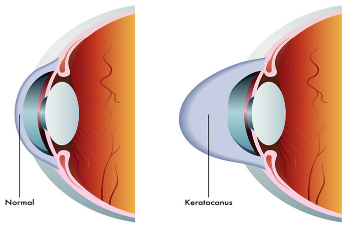
Keratoconus
Progressive thinning and forward protrusion of the cornea resulting in irregular and conical shape. Usually bilateral with early age onset. There are changes in refraction and the quality of the vision gets worse with time, despite correction. The situation typically occurs when your cornea gets thinner and gradually starts to bulge outward into a cone shape. This can result in blurred vision and sensitivity to light and glare.
Keratoconus affects both eyes and usually one eye is affected more than the other. Those between the ages of 10-25 and can progress slowly for 10 years or longer. There is certain familiar inheritance and no clear trigger to develop has been discovered. The collagen fibers become progressively weaker and get broken. Some point to personal biochemical factors.
Risk factors for developing keratoconus condition are as follows;
- Family history
- Excessive rubbing and squinting of eyes
- Having medical conditions like hay fever, asthma, and down syndrome

Diagnosis
A comprehensive eye examination will be performed after reviewing your family history and other medical conditions. Various tests are followed to diagnose the keratoconus.
Eye Refraction – A phoropter (a device that contains wheels of different lenses) or a retinoscope (hand-held instrument) is used to measure your eyes for vision problems and eyesight evaluation.
Slit-lamp Examination – A low powered microscope is used here to view your eye by directing a beam of light on the eye surface. The lights aids in observing the shape of your cornea and other potential problems.
Keratometry – Here also a circle of light is used to check the shape of your cornea and to measure the reflection of your eye.
Corneal mapping – This is a computerized photographic test, namely corneal tomography, and corneal topography, that records images to create a detailed shape of your corneal structure. This can also measure the thickness of your cornea and detect the early signs of keratoconus.
Intracorneal Ring Segments
In more advanced cases, we can implant fine artificial ring segments in the peripheral cornea, which helps flatten and regularize the central cornea, thereby improving the unaided vision due to less irregularity, less myopia and astigmatism. They also give more strength to the cornea. This procedure is painless, simple to perform and takes only 5 -10 minutes per eye. The segments are implanted in Intralase created corneal tunnels that match the shape of the segments.
Special Contact Lenses
Glasses can be used in early stages but normally they don´t provide good vision. Semisoft or RGP or Rose K contact lens, can help in mild to moderate cases. The quality of vision is better compared with glasses because they create a regular refractive surgery on the cornea. However, sometimes are difficult to adapt or wear, and they don’t stop the evolution.
Hard contact lenses are the first step in treating advanced keratoconus. Many people often find it uncomfortable but adjust to wearing them with time as they can provide excellent vision. But for those who do not prefer wearing these can go for piggyback lenses where a hard contact lens will be placed on top of a soft lens.
Hybrid lenses are another type that has been involved in increasing comfort. These lenses are equipped with a rigid center with a soft ring around to make it less discomfort for the wearer.
Scleral lenses are used for more advanced stages of keratoconus, where the lens does not rest in the cornea but sits on the white part of the eye and vaults over the cornea without touching it.

Phakic IOLs Implantation
Once we have stopped the evolution of the keratoconus, Phakic lenses (ICLs) can reduce the refractive error and astigmatism, thereby significantly improving uncorrected vision.
The lenses are made of plastic or silicone and are placed on the patient’s eye without removing the natural eye lens. The phakic lens is inserted through a small incision and placed in front or behind the iris (the colored part of the eye).
The lenses can focus the light correctly, providing a clear vision without having the need to wear glasses or contact lenses.
Cross-Linking (CXL)
It is a procedure to strength the cornea increasing the rigidity of the collagen fibers. Riboflavin eye drops are applied followed by exposure of the cornea to ultraviolet radiation. This causes cross-linking, stiffening the cornea to prevent further changes in the shape.The aim of CXL is to stop or, at least slow the progression of the keratoconus. Some cases even regress. It can be used in early cases.
Before starting the procedure, you will be given drops to numb your eyes. The specially formulated riboflavin (B2) will be put to the eyes, as it can help to absorb the light better. It will take around 30 minutes to soak the drops into the cornea.
After that, you will be laid back in a chair to start with the light. Typically a treatment takes around 60-90 minutes to complete.
Keratoplasty
Book An Appointment
Fill the form below or call/whatsApp +971 50 309 3131 to book an appointment.
Early treatment can prevent long-lasting consequences
